


Retinal vein occlusion
Understanding retinal vein occlusion1,2
Retinal vein occlusion (RVO) takes place when one or a few of the blood vessels in the eye become blocked.
This can result in a sudden, painless change in vision.
If you’re concerned about yourself or a loved one, talk to your doctor and scroll to find out more.
Here are some of the key terms we use when talking about retinal vein occlusion:
- Retinal
The light-sensitive part of the eye that becomes damaged in RVO. - Vein
A blood vessel that carries blood away from the eye. - Occlusion
A blockage in a blood vessel that stops the flow of blood in that area.
- Blood supply
The movement of blood to an area, bringing oxygen and nutrients to keep it healthy. - Central vision
The part of your vision you use to focus clearly on something directly in front of you, like reading a book. - Peripheral vision
The part of your vision you use to gather rough information about your surroundings, like navigating through a crowd.
Symptoms of RVO2
The main symptom of RVO is a sudden, painless change in vision. However, there are others that may also occur.
While symptoms may vary from person to person, they’re usually a combination of those shown below:
Blurred vision
The centre of your vision may become smudged or blurry.

Temporary loss of central vision
You may struggle to see things directly in front of you, making tasks like reading more difficult.

Central and peripheral visual disturbances
You may struggle to see things straight ahead or in your general surroundings.

Patches of vision loss
You may start to see ‘floating’ dark spots or lines in your vision.
RVO risk factors1,3,4
There are a number of risk factors to watch out for in RVO, including:

Age
RVO is most common in people aged 50 or over.

High blood pressure
This is also called hypertension.

Hardening of arteries
This is also called atherosclerosis.

All types of diabetes
Type 1, type 2, and gestational diabetes can all increase your risk of developing RVO.

Glaucoma
This is a condition that causes fluid to build up in the eye which can cause optic nerve damage.
How does RVO affect the eye?
Healthy eye


The retina, at the back of the eye, detects light. This information is sent to the brain to be processed as sight. In a healthy eye, the macula is the central part of the retina responsible for clear vision.3
Eye with RVO


This image is meant to show you one of the symptoms you may experience. Symptoms can vary depending on the severity level of the disease and will be unique for each person. If you are concerned about your vision, talk to your doctor.
An RVO blocks blood supply to one or a few of the blood vessels in the retina. This can affect the oxygen supply to the retina, damaging it and causing vision loss.
How does RVO develop?1,2,5
When blood supply is blocked to the retina it doesn't get enough oxygen.

As a result, the retina can’t send information to the brain to process vision.

This blockage can also cause swelling.
Without treatment, this can result in sudden and severe vision loss.
Two types of RVO1,2,5
There are two types of RVO, each depending on the type of blood vessel that’s affected.
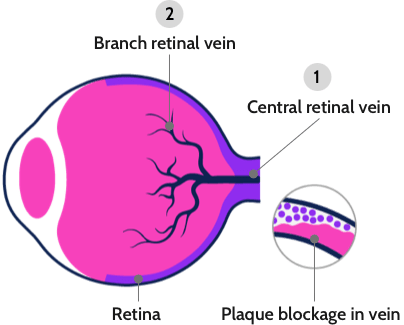
Branch RVO
When smaller veins, or branches, in the retina are blocked we experience branch RVO (BRVO).
Central RVO
When the main vein in the retina is blocked, we experience central RVO (CRVO). This type of blockage is usually the most serious of the two.
References
1. Macular Society. Retinal vein occlusion. [Internet; cited March 2021]. Available from: https:// www.macularsociety.org/retinal-vein-occlusion
2. MacDonald, D. (2013). The ABCs of RVO: A review of retinal venous occlusion. Clinical and Experimental Optometry, n/a. https://doi.org/10.1111/cxo.12120
3. Petr Kolar, "Risk Factors for Central and Branch Retinal Vein Occlusion: A Meta-Analysis of Published Clinical Data", Journal of Ophthalmology, vol. 2014, Article ID 724780, 5 pages, 2014. https://doi.org/10.1155/2014/724780
4. Laouri M, Chen E, Looman M, Gallagher M. The burden of disease of retinal vein occlusion: review of the literature. Eye. 2011; 25:981-8.
5. Retinal Vein Occlusion. Guy’s and St Thomas. [Internet; cited April 2021]. Available from: https://www.guysandstthomas.nhs.uk/resources/patient-information/eye/retinal-vein-occlusion.pdf